INTRODUCTION
The Elbow joint is considered to be modified (hinge variety of synovial joint). The Elbow joint consists of articulations between the distal humerus and the proximal radius and ulna. The function of the elbow joint is to extend and flex the arm grasp and reach for objects. The elbow joint is considered to be compound joint or hinge variety of synovial joint.
The Elbow joint has (one degree of freedom) which permits the motion of flexion and extension in sagittal plane around coronal axis. A small amount of axial rotation and abduction and adduction of ulna occurs during flexion and extension which is why it is considered to be a modified hinge joint.
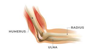
Image /Google
Table of Contents
The elbow joint includes the following joint-
- Humero-radial joint:- this joint is found between the articulating point of the capitulum, humerus and the head of the radius, which allows movements like flexion, extension supination and pronation.
- Humero-ulnar joint:- This joint is found between the articulating point of the trochlea on the medial aspect of humerus and trochlear notch on the proximal ulna.
- Proximal radio-ulnar:- This joint is found between the articulating point of the head of the radius and the radial notch of the ulna which allows to perform movements like supination and pronation.

Structures Of The Elbow Joint.
The proximally articulating surfaces are as follows:-
- The articulating surfaces on the anterior aspect of the distal humerus, trochlea and the spherical capitulum. These structures are situated between the medial and lateral epicondyles of the humerus. The trochlea forms the proximal portion of the medially located humeroulnar articulation and is set at an angle on the medial aspect of the distal humerus.
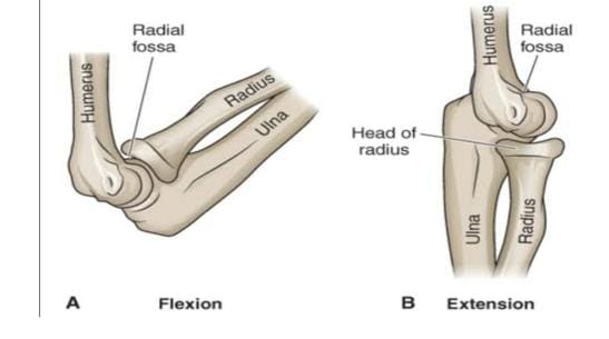
- The trochlear groove spiral obliquely around the trochlea and divides the trochlea into medial and lateral portion. The identation in the humerus called the coronoid fossa is designed to recieve the coronoid process of the ulna at the end of the elbow flexion range of motion (ROM). The radial fossa just above the capitulum is designed to receive the head of radius in full elbow flexion.
The distal articulating surfaces are as follows:-
- The ulnar articulating surface of the Humero-ulnar joint is a deep, semicircular concavity called the trochlear notch.
- The proximal portion of the notch is divided into two unequal parts by the trochlear ridge that corresponds to trochlear groove on the humerus. The distal articulating surface of the Humero-radial joints is composted of the head of the radius. The radial head has slightly cup-shaped concave surface called the fovea that is surrounded by a rim. The radial head’s convex rim fits into the capitulo-trochlear groove.
Joint Articulation
Motion at the Humero-ulnar joint occurs primarily as a sliding motion of the trochlear notch of the ulna on the trochlea of the humerus,
Image/Google
- In flexion, the trochlear notch of the ulna until the coronoid process of the ulna enters the coronoid fossa in full flexion. The articulating surface of the trochlea does not contact the deepest portion of the notch unless the joint is heavily loaded. The articulation between the radial head and the capitulum at the Humero-radial joint involves sliding the shallow concave radial head over the convex surface of the capitulum.
- The humeral capitulum is slightly smaller than the corresponding radial fovea, so the joint is incongruent (unsuitable).
In full flexion, the rim of the radial head slides into the radial fossa as the end of the flexion range is reached. - In full extension, no contact occurs between the articulating radial head and capitulum.
Function of the Joint Capsule.
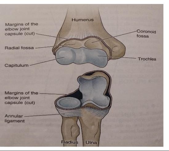
- The Humero-ulnar, Humero-radial and Proximal radio-ulnar joints are enclosed in a single joint capsule. The Capsule passes just below the annular ligament to attach to the posterior and inferior margins of the neck to the radius. The capsule is fairly large, weak in anteriorly and posteriorly which contains synovial folds that are able to allow wide range of motion while maintaining the stability of the Elbow Joint.
- Medially and laterally, the Capsule continues with the collateral ligaments. The proximal humeral attachment of the capsule is just the coronoid and radial fossa, whereas the capsule attaches distally to the ulna along the margin of the coronoid process and also attaches with the edge of the olecranon process.
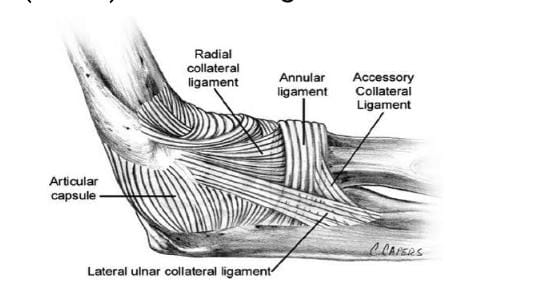
Ligaments of Elbow Joint.
The two main ligaments of elbow joints are as follows:-
1- Medial (ulnar) Collateral Ligament.
2- Lateral (radial) Collateral Ligament.
image/google
- Medial (Ulnar) Collateral Ligament:- The Medial Collateral Ligament is consist of the Anterior, Posterior and Transverse region. The anterior and posterior region is said to be as anterior and posterior Oblique ligament or Bundles. The Anterior bundle of the medial collateral ligament is considered to be primary ligamentous restraint to valgus stress from 20° to 120° of the elbow flexion.
- The Posterior bundle extends from the posterior aspect of the medial epicondyles of the humerus to attach to the coronoid and olecranon process of the ulna. The Posterior bundle limits elbow extension but plays a less significant role than the anterior bundle in providing valgus stability for the elbow.
- The Transverse fiber of Medial collateral ligament extend between olecranon and ulnar coronoid process. This ligament also known as Cooper’s ligament which appears to contribute little to valgus stability.
Functions of Medial collateral ligament.
- Stabilizes against valgus torques at the medial elbow.
- Limits extension at the end of the elbow extension ROM.
- Guide joint motion throughout flexion ROM.
- Provides some resistance to longitudinal distraction of the joint surfaces.
Lateral Collateral Ligament includes the following ligament:-
1-Lateral (radial) collateral ligament
2- Lateral (ulnar) collateral ligament
3- The annular ligament
- The Lateral Radial Collateral ligament is a fan-shaped structure that extends from the inferior aspect of the lateral epicondyle of humerus to merge with the annular ligament that encircles the head of the radius.
- The Lateral Ulnar Collateral Ligament originates from the lateral epicondyle of the Humerus and insertion goes to supinator crest of the Ulna. It helps Ulna from rotating away from the trochlea.
- The Annular Ligament is a strong circular shape ligament which forms a ring around the radial head. It originates from the sigmoid notch of Ulna and insertion goes to head of the Radius. It’s main function is to stabilize the Radioulnar Joint.
Function of lateral collateral ligament:-
1.Stablizes elbow against varus torque.
2.Stablizes against combined varus and supination torques.
3.Stablizes radial head, thus providing a stable base for rotation.
4.Prevents subluxation of humeroulnar joint by securing ulna to humerus
5.Prevents the forearm form rotating off the humerus in valgus and supination during flexion from the fully extended position.
6.Maintains the stability of posterolateral rotation.
What is Carrying Angle?
Carrying angle is an acute angle between the long axis of the extended forearm and the arm. The normal carrying angle of the elbow joint is Males- 5° to 10° and in Female- 10° to 15°.
Know more-
To Learn and understand more about Infrared Radiation therapy click the given link https://physiocontent.com/infrared-radiation-therapy/
To learn and understand more about Relaxation Technique click the given link https://physiocontent.com/muscle-relaxation-techniques-and-benefits-of-relaxed-muscles/